Blog Roll: (Contributors)
» Hulet Smith, OT
» Megan Smith, PT
» Mike Price, OT
Topics:
Adaptive Devices
Adult Tricycles
Air Purifier
Allergy
Alternative Communication Devices
Alzheimer's Dementia Products
Aquatic Products
Arthritis Relief Products
Autism
Back Relief
Ball Pit/Pool
Bariatric
Bath Benches
Bathtub Lift
Bed Rails
Bedsores / Decubitus
Bidet
Body Solid Exercise Products
Breast Feeding Products
Bushel Trucks
Cancer
Catheters
CEU
Changing Bench
Child Car Seats
Child Care Products
Christmas Gifts
Clinic/Medical Equipment
Clinical Furniture
Cold and Flu
Communication Devices
Compression Garments
Computer Products
CPAP
Crutches
Daily Assistance Products
Daylight Lamps
Dental Care
Diabetes
Doctor's Office
Dysphagia
Electrodes
Electrolarynx
Emergency Preparedness
Ergonomic Equipment
Exam Tables
Exercise Products
Eyecare
Family Tricycles
First Aid Kits
Floor Scales
Fluidotherapy
Foot Drop
Foul Weather Gear
Furniture
Gait Trainers
General Articles
General Posts
Glassless Mirrors
Hand Sanitizer
Head Protection Helmets
Hearing Impaired
Heart Health
Heating Pad
Hip Fractures
Home Assistance Products
Home/Office Assistance
Hospital Beds
Hoyer Lifts
Hyperbaric chamber
Hyperthermia/Hypothermia
Ice/Hydration Carts
Impotence Products
Incontinence Products
Infection Control Gowns
Inspirational Stories
Lift Chairs
Light Therapy
Low Vision Products
Massage Tables & Chairs
Massage Units
Maternity
Medical Facility Products
Medical Scales
Multi-Sensory Environment
Natural Healing
Nebulizers
Non-Hospital Bedding
Nutritional Supplements
Office Furniture
One-Handed Products
Operating Room Devices
Ostomy Products
Oxygen Compressors
Oxygen Concentrator
Oxygen/Nebulizer Masks
Pain Relief
Paraffin Unit
Patient Lift
Patient Lifts
Patient Restraints
Patient Transfer Systems
Pediatric Bath Chairs
Pediatric Furniture
Pediatric Learning
Pediatric Recreation
Personal Listening Devices
Personal Warming Products
Physical Therapy
Pill Organizers
Pillows
Playground Equipment
Pool Lifts
Press Releases
Procedure Chairs
Pulse Oximeter
Reading Assistance
Reference Materials
Rehab Equipment
Rehabmart News
Rehabmart Newsletter
Respiratory Health
Rollators
Saunas
Scooters
Seniors
Shower Chairs
Shower Commode Chairs
Shower Gurney
Showers Chairs
Side Access Bathtubs
Skin Tear
Special Needs Dinnerware
Special Needs Seating
Special Report Articles
Splints
Sport Injuries
Standers
Staying Home
Stethoscopes
Stimulus Reward Toys
Stress Relief
Stroke
Strollers
Summertime Products and Summertime Fun
Talking Products
Therapy Balls
TheraTogs
Thermometers
Traction Devices & Tables
Treatment Tables
Ultrasound
Vibroacoustic Therapy
Vision Products
Walk-In Bathtub
Walking Aids
Walking Boot
Weighted Wearables
Wheelchair Accessories
Wheelchair Cushions
Wheelchair Lifts
Wheelchair Ramps
Wheelchair Transfer Systems
Wheelchairs
Women's Health
Work Hardening Products
Wound Care
System provided by Maximtech.com
Models Provide Visual References for Learning Anatomy
Posted in Reference Materials by Hulet Smith, OT on 9/20/2012
Whether one studies conventional medicine or a complimentary discipline such as massage or acupressure, a working knowledge of anatomy is part of the curriculum. While many classes use preserved cadavers to teach about the human body, models provide visual references in situations where cadavers may not be available or may not be suitable for the students in question, such as teaching elementary school children the basics of anatomy. They may also be used in clinics and private doctors' offices for patient education as well. Here are five model options from Rehabmart that offer anatomical education for both the teacher and the student.

At 33 inches tall, the Miniature Skeleton is a valuable classroom tool for introducing elementary and junior high students to the skeletal system. It has a removable calavarium and is mounted by the pelvis to the display stand. For reference, a key card naming the individual bones is provided.

For more advanced students such as those studying medicine or some form of therapy, the Standard Skeleton is a life-sized replica of the human skeleton. The arms and legs are removable and the skull can be divided into three sections. It can be used to demonstrate physical therapy maneuvers. The Standard Skeleton comes with a dust cover and is mounted by its pelvis onto a base with casters for easy transport around the classroom or from one room to another.

In addition to removable limbs and skull, the Deluxe Skeleton has a flexible spine. Enhancing the learning experience even further are painted-on depictions of the muscle origins, nerve endings, and insertions on its left side while the right side features numbers for identifying bones, bone parts, fissures, and foramen on the left. Like the Standard Skeleton, it comes with a rolling base for easy transport, is pelvic-mounted to the base, and has a dust cover.
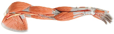
Rehabmart offers two options for detailed study of the upper limbs. The first is the Full Arm Model, which allows students to see the anatomical details of the arm in all three dimensions. The Full Arm Model is two feet long, and comes with a detachable stand. It's made of colorful plastic to enhance its realistic appearance. Five parts can be removed to allow for closer examination of the nerves and blood vessels as well as helping students and patients visualize how the muscles fit and work in conjunction with one another.

The second option is the Muscles of the Hand Model. It's made of latex and like the Full Arm Model, it has been colored to give it a realistic appearance. It comes with five detachable parts, including a removable palmar aponeurosis, allowing a student to see clearly the hand's anatomy layer by layer. Each nerve, blood vessel, and muscle has been labeled for easy identification. The model provides an illustration of the carpal tunnel as well as the nerves and bones of the hands. A stand is included.
Whether it's in a classroom or in a doctor's office, any of these models will enhance the patient's or student's understanding of the human body and how it works.
Fran Jablway,
Medical Consumer Writer
and
Hulet Smith, OT
Rehabmart Team Leader & CEO